“My time in the Dean lab allowed me to develop a deeper understanding into how ideas are developed in the lab and how much collaboration is involved in research.” – Lauren
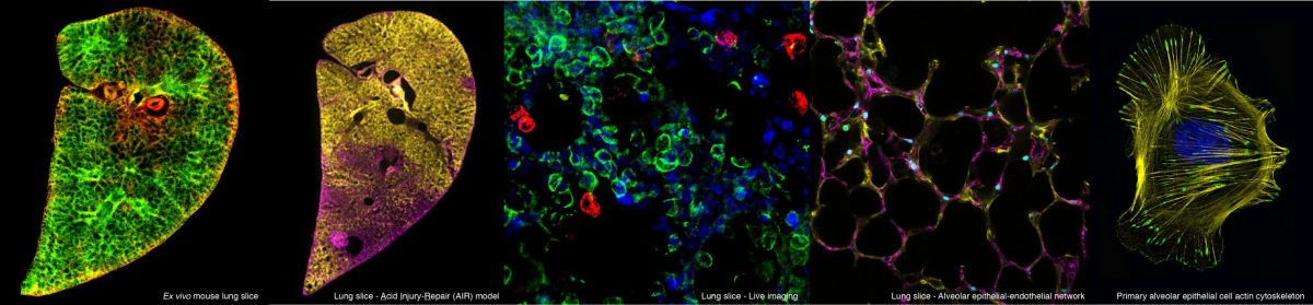
“For my master’s thesis I was lucky enough to undertake a 6-month research project with the dean lab. This experience was incredible, for the first time I was truly integrated into a labs’ research. For my project I was able to work with the on the Acid Injury Repair (AIR) model. This novel model uses hydrochloric acid to mimic lung injury and allows for the study of lung repair and regeneration. Working with Rosin Mongey, who established the AIR Model, I tested ways to image dynamic processes happening in the injured tissue.
Throughout my project I learnt new techniques and concepts. Thanks to the great supervision of Rosin and the FILM department, I became confident with microscopy, an area I knew little about. As microscopy is a central technique in biological research, these newly developed skills will help me in future research.
Alongside my lab work, I was invited to attend weekly lab and section meetings. These meetings helped to highlight the collaborative nature of research. In lab meetings I was kept up to date with the labs work and was able to present and receive feedback on my own, whilst in section meetings, I learnt about the research being performed by other groups.
My time in the Dean lab allowed me to develop a deeper understanding into how ideas are developed in the lab and how much collaboration is involved in research. I hope that the skills I have learnt during this project will help me in a future research career.”
– Lauren
 :
: