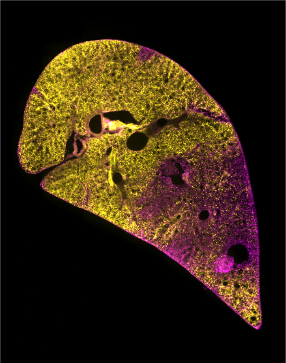
A lung slice showing isolated areas of damaged (purple) and undamaged (yellow) tissue
Studying the mechanisms of lung repair and regeneration in the human lungs is challenging given their critical role. We have developed a new model, using slices of lung tissue, that can be used to study lung repair and regeneration. The Acid Injury and Repair (AIR) model works by using hydrochloric acid to injure a small part of the tissue slice whilst the surrounding area remains uninjured. This mimics the pattern of injury often observed in lung diseases. The AIR model enables tracking of different cell types, including stem cells as well as providing a platform to test potential new treatments to repair the lungs.
Our new research article “The Planar Polarity Component VANGL2 Is a Key Regulator of Mechanosignaling” has been published in Frontiers in Cell and Developmental Biology.
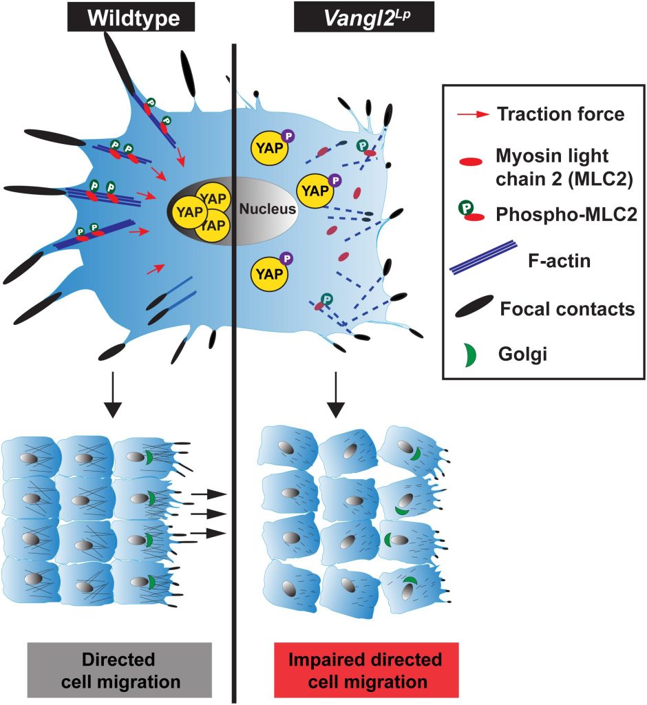
In this study, Dr Sek Shir Cheong discovers how a mutation in the VANGL2 gene (Vangl2Lp), in mutant mice, interferes with normal mechanosignalling and directed cell migration which contribute to abnormal formation of alveoli, the site of gas exchange in the lungs. Mechanosignalling, “mechano-signalling”, simply put, is a signalling process induced by mechanical forces.
By monitoring in real time the movement of alveolar epithelial cells (an important cell type that form alveoli) in slices of lung tissue, we found that cells from the Vangl2Lp/+mutant mice had slower and more restricted movement compared to cells from normal healthy mice. This resulted in fewer alveoli being formed, which reduces lung function in the mutant mice. To help us understand what causes the abnormal movement in these mutant cells, we isolated epithelial cells from the tracheas and lungs of Vangl2Lp/+ mutant mice, and labelled different components in the cells using antibodies. This process revealed that the Vangl2Lp mutation caused disruption of the actomyosin network (a kind of scaffold present in cells that is analogous to the musculoskeletal system in the human body), as well as focal adhesions, which function as molecular clutches, enabling cells to attach to their surrounding matrix. In addition we found a reduction in key proteins responsible for mechanosignalling within the lung epithelial cells. All of these abnormalities leads to the the cells being floppy as they are unable to generate force, thereby affecting their ability to move or migrate which is particularly important during the process of lung formation or lung repair after injury.
Lastly, we tested a drug WNT5A, a molecule that belongs to the same pathway as VANGL2, and showed that it could improve wound healing ability in the mutant cells, suggesting that WNT5A could be a potential target for lung repair.
This article highlights a previously unknown role of VANGL2 in command and control of mechanosignalling.
Comparison of cellular mechanics in wildtype and Vangl2Lp cells.
During my second year of medical school I decided that I wanted to gain some lab experience over Summer, as I realised medical research was an area I’d like to explore further. I was very fortunate to arrange to spend my Summer in the Dean lab and subsequently worked with Charlotte and Matt to design a project that would contribute to the labs work in investigating the mechanisms underlying Congenital Pulmonary Airway Malformation (CPAM). I applied for and gratefully received a Wellcome Student Scholarship to facilitate this, producing a report based on my findings. I found it a fantastic experience working within the research team and gained valuable insight into clinical academia through collaborating with clinicians at the Royal Brompton Hospital.
I have subsequently presented the results of my project at the 2019 Pathology Society Winter meeting held at the Royal Society of Medicine, and this year our article was published in BMJ Thorax. DOI: 10.1136/thoraxjnl-2020-214752.
My experience in the Dean lab has been integral to informing my choice to pursue academic research alongside my clinical work. I have since completed my BSc at Imperial in Cardiovascular Sciences, during which I conducted a lab project at Tokyo Medical and Dental University, and I am currently working as an academic foundation doctor in which I will complete my academic rotation in transplant and regeneration at Addenbrooke’s hospital, Cambridge.
Taylor B et al. Mechanism of lung development in the aetiology of adult congenital pulmonary airway malformations. Thorax 2020
A new study cross-disciplinary study from several groups in NHLI demonstrates the importance of lung development genes in regulating adult lung function. This project used UK Biobank to investigate whether lung development genes influence adult lung function. Future experimental investigation of these developmental pathways could lead to druggable targets to improve lung function.
Am J Respir Crit Care Med. 2020 May 11. doi: 10.1164/rccm.201912-2338OC. Online ahead of print.PMID: 32392078
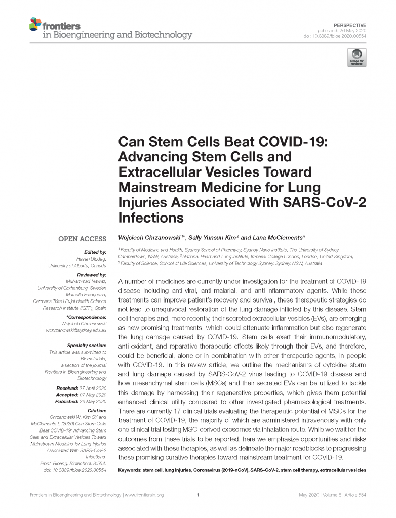
A new paper from Sally Kim and colleagues looks at the potential for stem cells and Extracellular vesicles as potential treatments for lung damage caused by COVID-19.
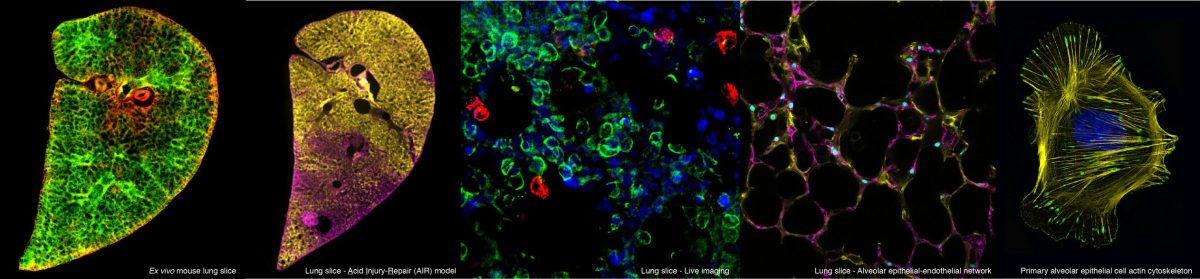
Septation, cell clustering and epithelial cell migration of epithelial cells to existing airspaces in P3 PCLS. Epithelial cell dynamics in P3 PCLS. EpCAM-FITC (green) and SiR-DNA (magenta) labelled P3 PCLS imaged for 12 hours 45 minutes at 15 minute intervals. Red arrows indicate migrating septa, as one existing airway subdivides into two (a1-a2 and a3-a4). Blue circles indicate areas where cell clustering can be seen. Migrating epithelial cells intercalate with existing alveolar wall epithelial cells (yellow arrows) around two airspace, a5 and a6.Epithelial cells integrate into an existing airspace EpCAM-FITC (red) and SiR-DNA (cyan) labelled P3 PCLS imaged for 14 hours 15 minutes at 15 minute intervals. Epithelial cells 1,2 and 3 (green arrows) migrate towards an existing alveolar airspace (white circle).Visualisation of the epithelium and capillary network in P3 PCLS. EpCAM-FITC (green) and PECAM-Alexa 647 (red) labelled P3 PCLS imaged for 12 hours 30 minutes at 15 minute intervals. Both EpCAM positive epithelial and PECAM positive endothelial cells can be seen in an extending septum during septation.Control and blebbistatin treated P3 PCLS EpCAM-FITC (green) and SiR-DNA (magenta) labelled P3 PCLS treated with DMSO control media (A), imaged for 14 hours at 15 min intervals or 50µM blebbistatin containing media (B), imaged for 14 hours 15 minute intervals. a= airspaces.
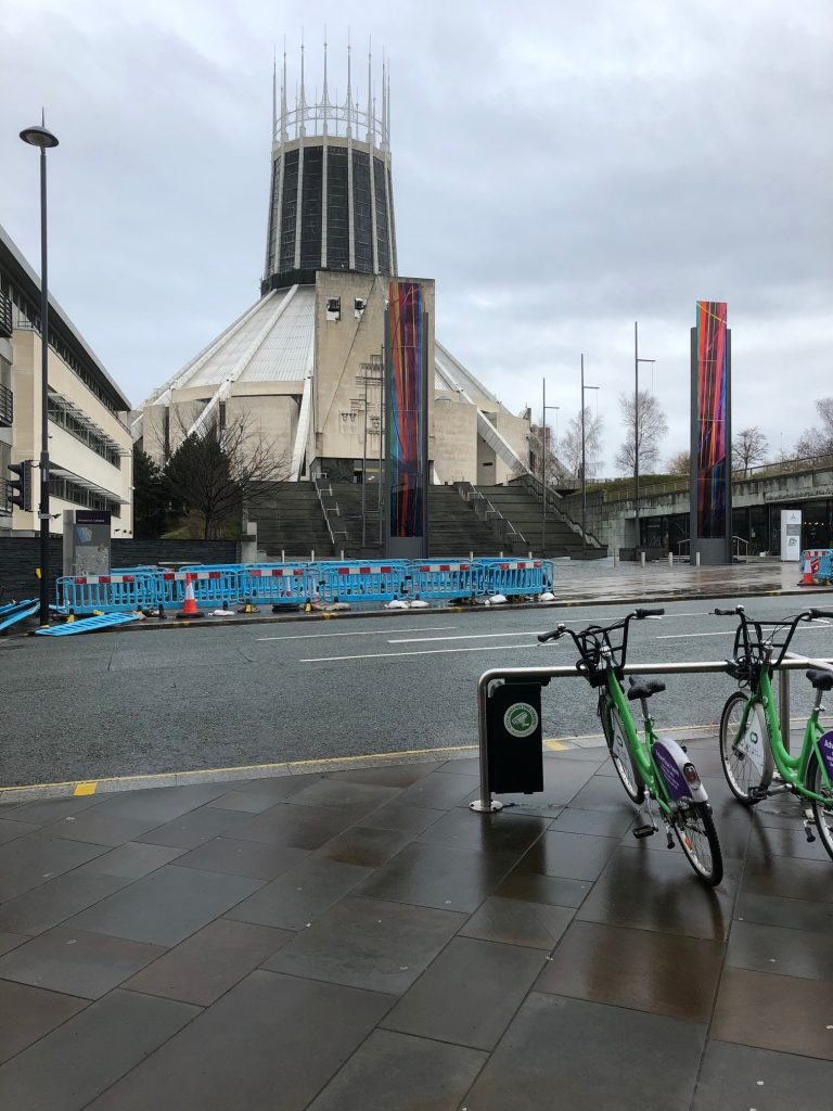
On 29th February Charlotte attended the 5th Annual National Liverpool Research Conference. This student led initiative is a one day meeting specifically aimed to encourage medical students to get involved in research. The day began with several talks by senior clinical and basic research scientists on a variety of topics such as ‘engineering an artificial womb’ and Charlotte’s talk ‘towards lung regeneration:tools and mechanisms’. There was also a poster and oral presentation competition, which showcased the many different projects that students have been working on. Participants were also able to attend workshops to learn more about routes into clinical academic research. A huge thanks to all the organisers and attendees for a thoroughly enjoyable day.
Meeting was held at the Liverpool Medical Institute opposite the catholic cathedral (pictured).
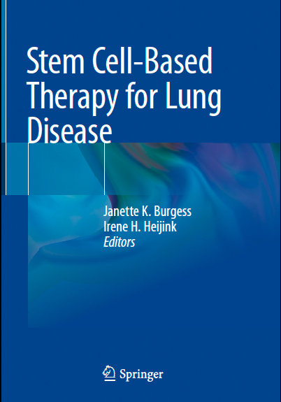
For a really usefull up to date look at stem-cell based therapies, take a look at this book which has just been published. The chapter on ‘stem cell delivery systems and devices’ was co-authored by Sally Kim, a current ERS/EMBO fellow in the lab.
Sek Shir recently presented her interesting new data at a poster entitled “Disruption of The Planar Cell Polarity (PCP) Component Vangl2 Alters Cell Mechanics in Loop-tail Mice” at the EMBO/EMBL symposium in Heidelberg, Germany between 3-6 July, 2019.
The symposium aims at bringing together world-leading experts in the fields of mechanobiology, cell biology and developmental biology studying the mechanical basis of cell and tissue morphogenesis. Particular emphasis was given to quantitative approaches analysing how force production, transduction and reception drives cell and tissue morphogenesis from the molecular scale to the organismal scale. The Symposium aims to provide a comprehensive overview of both experimental and theoretical advances providing insight into the molecular, cellular and biophysical mechanisms by which cells, tissues and entire organisms take shape.