Investigating mechanisms of lung tissue regeneration
Real-time imaging of lung slices
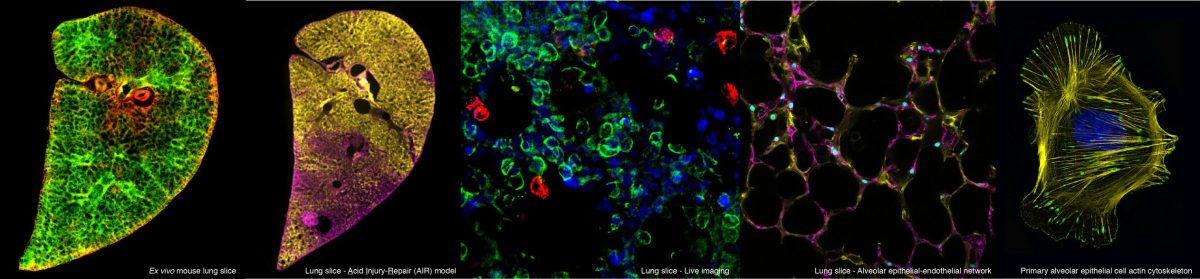
Septation, cell clustering and epithelial cell migration of epithelial cells to existing airspaces in P3 PCLS. Epithelial cell dynamics in P3 PCLS. EpCAM-FITC (green) and SiR-DNA (magenta) labelled P3 PCLS imaged for 12 hours 45 minutes at 15 minute intervals. Red arrows indicate migrating septa, as one existing airway subdivides into two (a1-a2 and a3-a4). Blue circles indicate areas where cell clustering can be seen. Migrating epithelial cells intercalate with existing alveolar wall epithelial cells (yellow arrows) around two airspace, a5 and a6.Epithelial cells integrate into an existing airspace EpCAM-FITC (red) and SiR-DNA (cyan) labelled P3 PCLS imaged for 14 hours 15 minutes at 15 minute intervals. Epithelial cells 1,2 and 3 (green arrows) migrate towards an existing alveolar airspace (white circle).Visualisation of the epithelium and capillary network in P3 PCLS. EpCAM-FITC (green) and PECAM-Alexa 647 (red) labelled P3 PCLS imaged for 12 hours 30 minutes at 15 minute intervals. Both EpCAM positive epithelial and PECAM positive endothelial cells can be seen in an extending septum during septation.Control and blebbistatin treated P3 PCLS EpCAM-FITC (green) and SiR-DNA (magenta) labelled P3 PCLS treated with DMSO control media (A), imaged for 14 hours at 15 min intervals or 50µM blebbistatin containing media (B), imaged for 14 hours 15 minute intervals. a= airspaces.