Developmental signalling in lung disease and repair
• Wnt/PCP signalling • Retinoic acid signalling • Shh signalling
Many signalling pathways that regulate lung development are also important for adult lung repair. We are investigating how these pathways influence tissue regeneration and whether they can be manipulated to enhance lung repair.
› WNT/PCP signalling
We have shown that the Wnt/planar cell polarity (PCP) pathway plays a key role in adult lung homeostasis and repair . WNT5A enhances the alveolar epithelial progenitor response following injury, while disruption of PCP signalling affects epithelial cell behaviour and repair. We are continuing to investigate how this pathway can be modulated to promote effective lung regeneration.
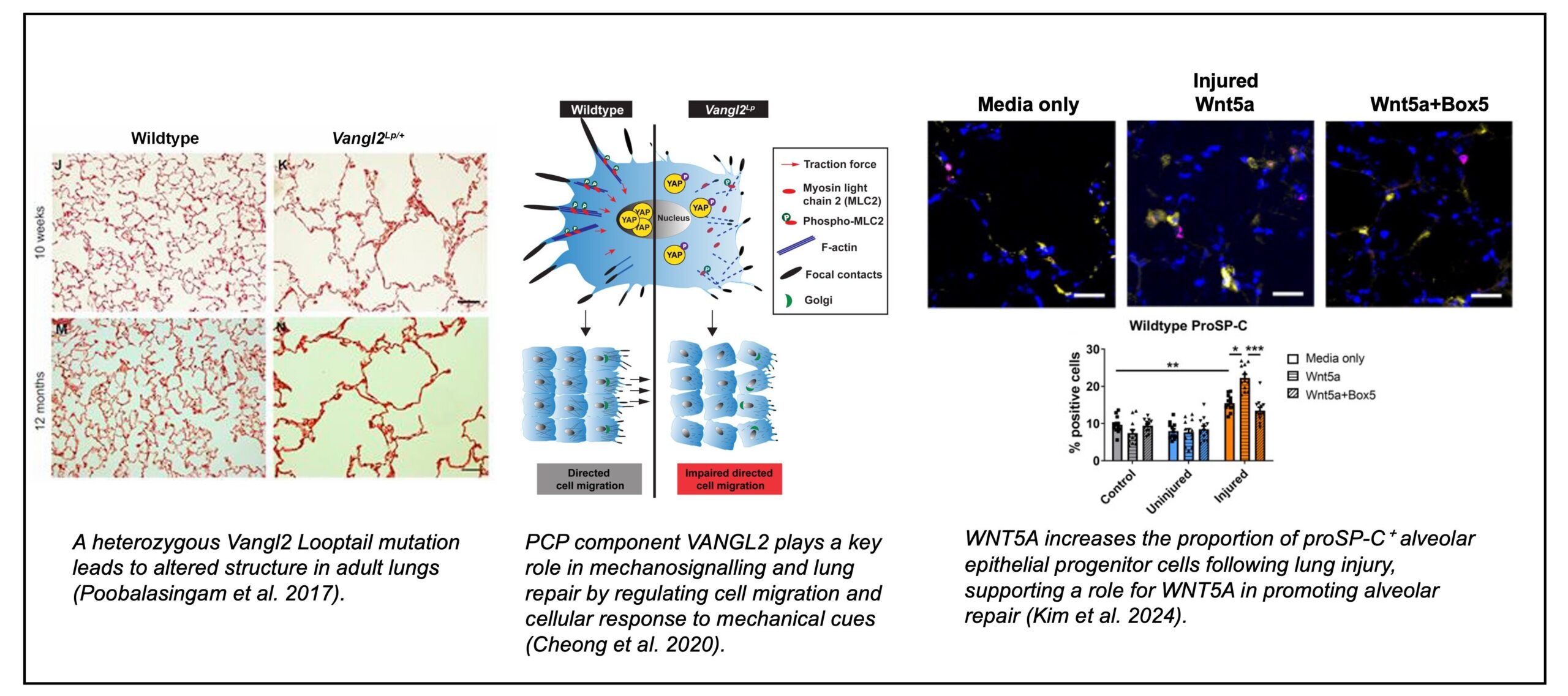
 Related Publications: Kim et al. 2024 Cheong et al. 2020 Poobalasingam et al. 2017
› Retinoic acid (RA) signalling
Our work has identified a role for retinoic acid (RA) in promoting endothelial angiogenesis and enhances paracrine signalling to alveolar epithelial cells, with TGFα/EGFR signalling contributing to epithelial repair.
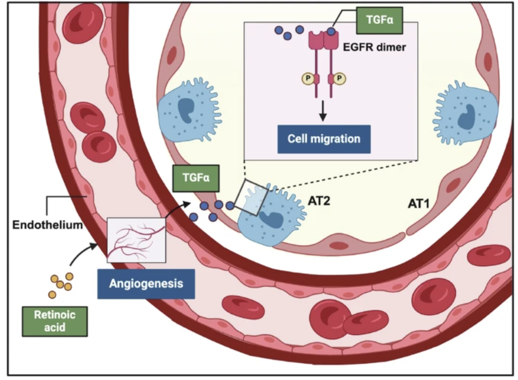
Related Publication: Cheong et al. 2026
› Other developmental pathways / targets
We are also investigating the role of WNT11, a non-canonical Wnt ligand, and Sonic Hedgehog (Shh) signalling in lung injury and repair, with the aim of understanding how these pathways regulate tissue regeneration and identifying potential therapeutic strategies to enhance lung repair.
Real-time imaging of lung development and adult lung repair
• Postnatal alveologenesis • Adult lung repair
Real-time imaging enables us to discover details about the role of genes in lung development that would not have been possible using 2D/static imaging techniques. We have established Precision-Cut Lung Slice models from human and mouse tissue, future work will continue to use this important pre-clinical tool.
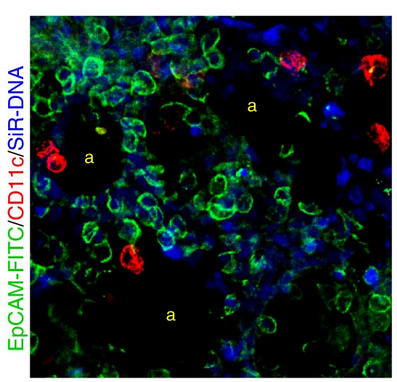
›Real-time imaging of postnatal PCLS during alveolarisation
Alveoli, the gas-exchanging compartment of the lungs, are thought to form by cells repeatedly sub-dividing airspaces eventually creating the large surface area needed for respiration. Despite their critical function, current knowledge is based solely on 2D pictures. We have used live 3D imaging of alveolarisation in slices to determine precisely how alveoli form.
Click here to view movies generated from real-time imaging of lung slices.
Related publications: Akram et al. 2019 Akram et al. 2019
› Real-time imaging of adult PCLS during lung repair
We also use this system to evaluate potential therapies for enhancing adult lung repair. Cell-specific markers enable us to track the dynamic movement and behaviour of different cell populations in real time following injury.
Click here to view the protocol of the Acid Injury and Repair (AIR) model
Real-time imaging of alveolar type II (ATII) epithelial cells in adult PCLS. ATII cells are labelled with MHCII-Alexa Fluor 594 (red) in an uninjured region adjacent to an acid-injured region. The cell membrane is shown in green. Images were acquired every 30 minutes for 19.5 hours, from 48-68 h post-injury, capturing dynamic ATII cell behaviours during the early response to tissue injury.
Lung-derived extracellular vesicles and lung repair
We are investigating how the physicochemical and biological composition of extracellular vesicles (EVs) are altered across lung development and ageing. EVs are heterogeneous particles (50 – 1000 nm) secreted by cells that share biological information between cells through encapsulated signalling proteins, nucleic acids and lipids. Research into EV biology has grown exponentially since their potential as non-cellular tissue modifiers has begun to be realised. We are currently addressing a critical gap in the knowledge about EV biology using a variety of tools and techniques previously established in the lab: precision-cut lung slices, the acid injury and repair (AIR) model, fluorescence microscopy, and atomic force microscopy.
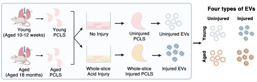
Using EVs isolated from mouse precision-cut lung slices (PCLS), we are comparing EVs derived from postnatal and adult lungs, as well as from adult and aged lungs, to identify age-dependent differences in their biological activity and molecular cargo.
Our work indicates that EVs from different stages of lung development and ageing have distinct effects on lung repair, with young lung-derived EVs promoting repair responses in injured tissue. We are further investigating the molecular signals carried by these EVs to understand how they regulate tissue regeneration and whether they could provide new therapeutic strategies for promoting repair in the ageing lung.
Related publication: Chen et al. 2026