Investigating mechanisms of lung tissue regeneration
Real-time imaging of Precision-Cut Lung Slices (PCLS)
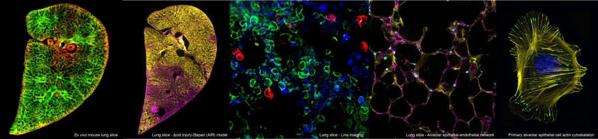
We have established advanced real-time live 3D imaging of precision-cut lung slices (PCLS), enabling dynamic cellular behaviours and tissue remodelling to be visualised in real time. Unlike conventional 2D or static imaging, this approach allows us to directly observe how cells move, interact and reorganise within the native lung tissue architecture. The detailed real-time imaging of PCLS setup has been published and can be found here.
Our real-time imaging platform can be used to investigate two key processes:
1. Postnatal alveolarisation We established a novel live-imaging approach that enabled us to visualise postnatal alveolarisation in PCLS for the first time. By following alveolar development over time, we identified previously unrecognised cellular behaviours and strategies used by epithelial cells to form new alveolar structures (Akram et al. 2019).
Septation, cell clustering and epithelial cell migration of epithelial cells to existing airspaces in P3 PCLS. Epithelial cell dynamics in P3 PCLS. EpCAM-FITC (green) and SiR-DNA (magenta) labelled P3 PCLS imaged for 12 hours 45 minutes at 15 minute intervals. Red arrows indicate migrating septa, as one existing airway subdivides into two (a1-a2 and a3-a4). Blue circles indicate areas where cell clustering can be seen. Migrating epithelial cells intercalate with existing alveolar wall epithelial cells (yellow arrows) around two airspace, a5 and a6.Epithelial cells integrate into an existing airspace EpCAM-FITC (red) and SiR-DNA (cyan) labelled P3 PCLS imaged for 14 hours 15 minutes at 15 minute intervals. Epithelial cells 1,2 and 3 (green arrows) migrate towards an existing alveolar airspace (white circle). Visualisation of the epithelium and capillary network in P3 PCLS. EpCAM-FITC (green) and PECAM-Alexa 647 (red) labelled P3 PCLS imaged for 12 hours 30 minutes at 15 minute intervals. Both EpCAM positive epithelial and PECAM positive endothelial cells can be seen in an extending septum during septation.
2. Cell behaviour during adult lung repair We have also extended this approach to adult PCLS following lung injury, enabling us to track cell movement and behaviour in real time during the repair process. This provides a unique opportunity to investigate how cells respond to injury, migrate within the tissue and contribute to regeneration and repair.
Real-time imaging of alveolar type II (ATII) epithelial cells in adult PCLS. ATII cells are labelled with MHCII-Alexa Fluor 594 (red) in an uninjured region adjacent to an acid-injured region. The cell membrane is shown in green. Images were acquired every 30 minutes for 19.5 hours, from 48-68 h post-injury, capturing dynamic ATII cell behaviours during the early response to tissue injury.
Together, these capabilities provide a powerful platform for understanding the dynamic processes that shape lung development and repair, and for identifying potential therapeutic strategies to enhance lung regeneration.